
|

|
Data Sheet
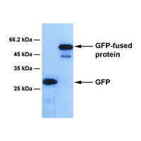
BackgroundGreen fluorescent protein (GFP) was first isolated from jellyfish Aequorea victoria, and has been used as a reporter or a tag of fusion protein for expression analysis in molecular and cell biology. Different mutants of GFP have been engineered to produce varieties of colors such as blue (BFP), cyan (CFP) and yellow (YFP). These color mutants provide powerful tools for fluorescence microscopy studies as they are less harmful in living cells, and in vivo imaging as they are heritable and can be expressed throughout an animal. In the presence of long-wave UV light, GFP emits green light, which has a major excitation peak at a wavelength of 395 nm. Studies have shown that GFP is an exceptionally stable protein in a wide range of pH and temperature. SourceThis is a mouse monoclonal antibody, which was raised against the full-length GFP protein. Gene SymbolGFP (A. victoria) IsotypeIgG2b Physical FormFreeze-dried powder from 1 × PBS solution SpecificityThis antibody detects GFP and its fusion proteins. Other color mutants have not been tested Molecular Weight27 kDa (GFP) ApplicationWestern blotting (WB, dilution range: 1:1,000 - 10,000). Other applications have not been tested yet. StorageStore freeze-dried powder at 2 - 8°C upon arrival. When ready to use, rehydrate with 0.1 ml dH2O and centrifuge if not clear. For long-term storage, make aliquots and keep them at -20°C or below. Avoid repeated freezing and thawing cycles. Data>> Western blotting: HT-1080 cell extracts expressing GFP and its fusion protein prepared in 1% Triton-X lysis buffer.
Important NoteThis product is intended for research use only, not for use in human therapeutic or diagnostic procedures. |

|
Copyright © Lanleys International Inc. All rights reserved.
|
